What to expect
The GAE procedure, step by step
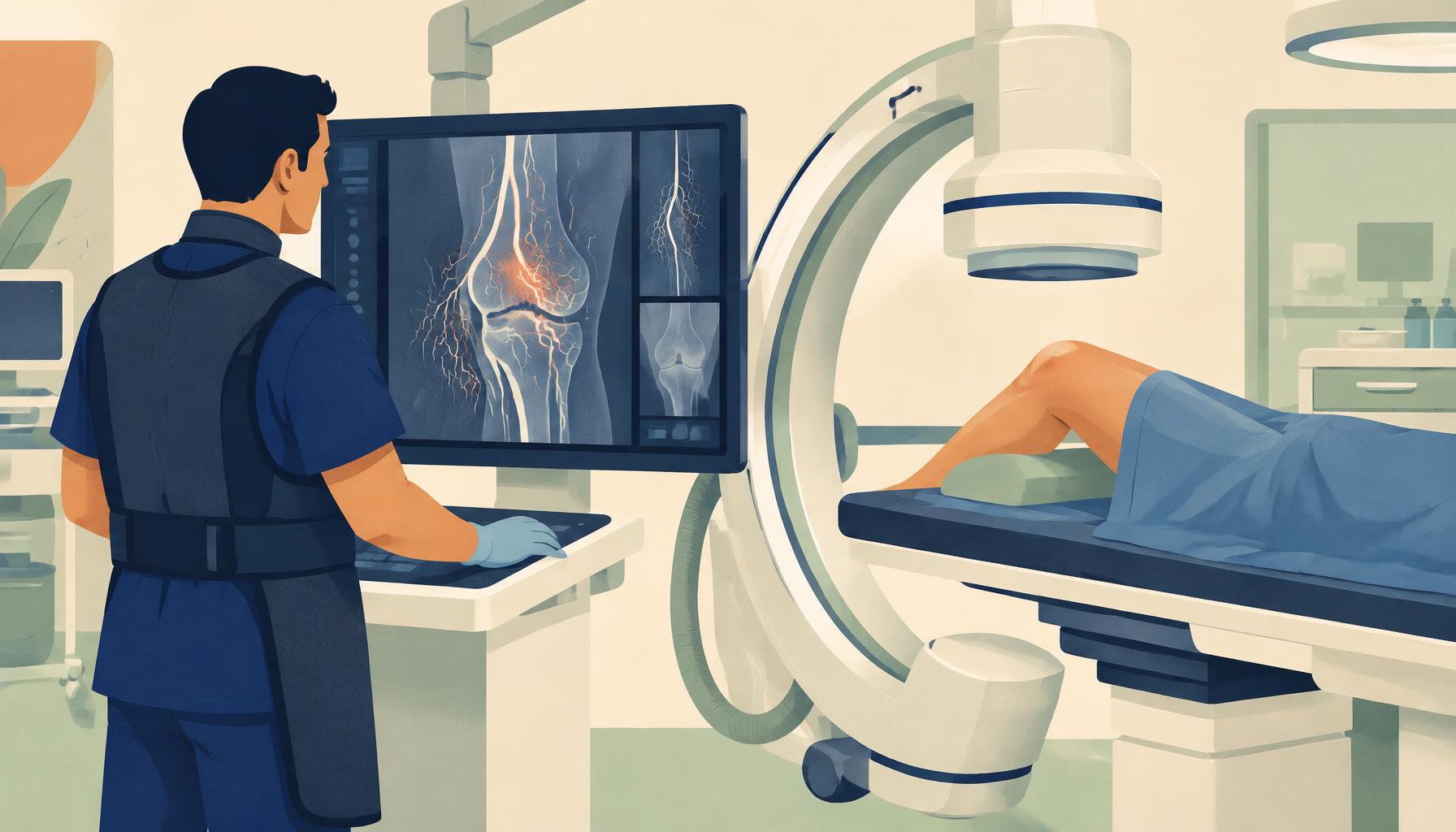
Step by step
- 1
Consultation & imaging
Exam, review of symptoms and X-rays; MRI may be ordered to assess synovitis and exclude other causes of pain.
- 2
Arrival & sedation
You arrive the day of the procedure, an IV is placed, and you receive moderate 'twilight' sedation — relaxed and comfortable, but breathing on your own.
- 3
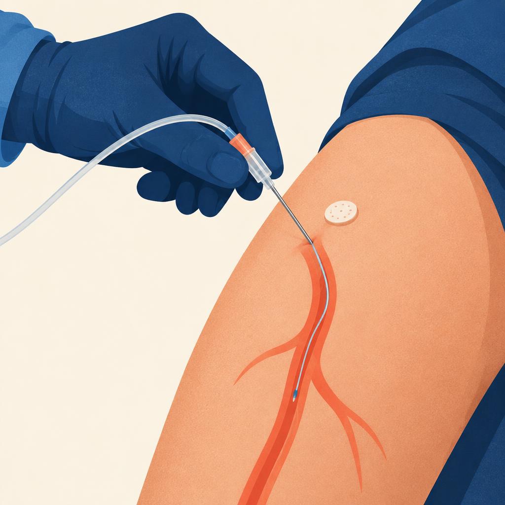
Pinhole access
After numbing the skin, a tiny needle puncture is made in an artery at the top of the thigh (or sometimes near the knee). No incision or stitches.
- 4
Mapping the genicular arteries
A thin catheter is guided under X-ray to the knee. Contrast dye maps the genicular arteries and highlights abnormal blush.
- 5
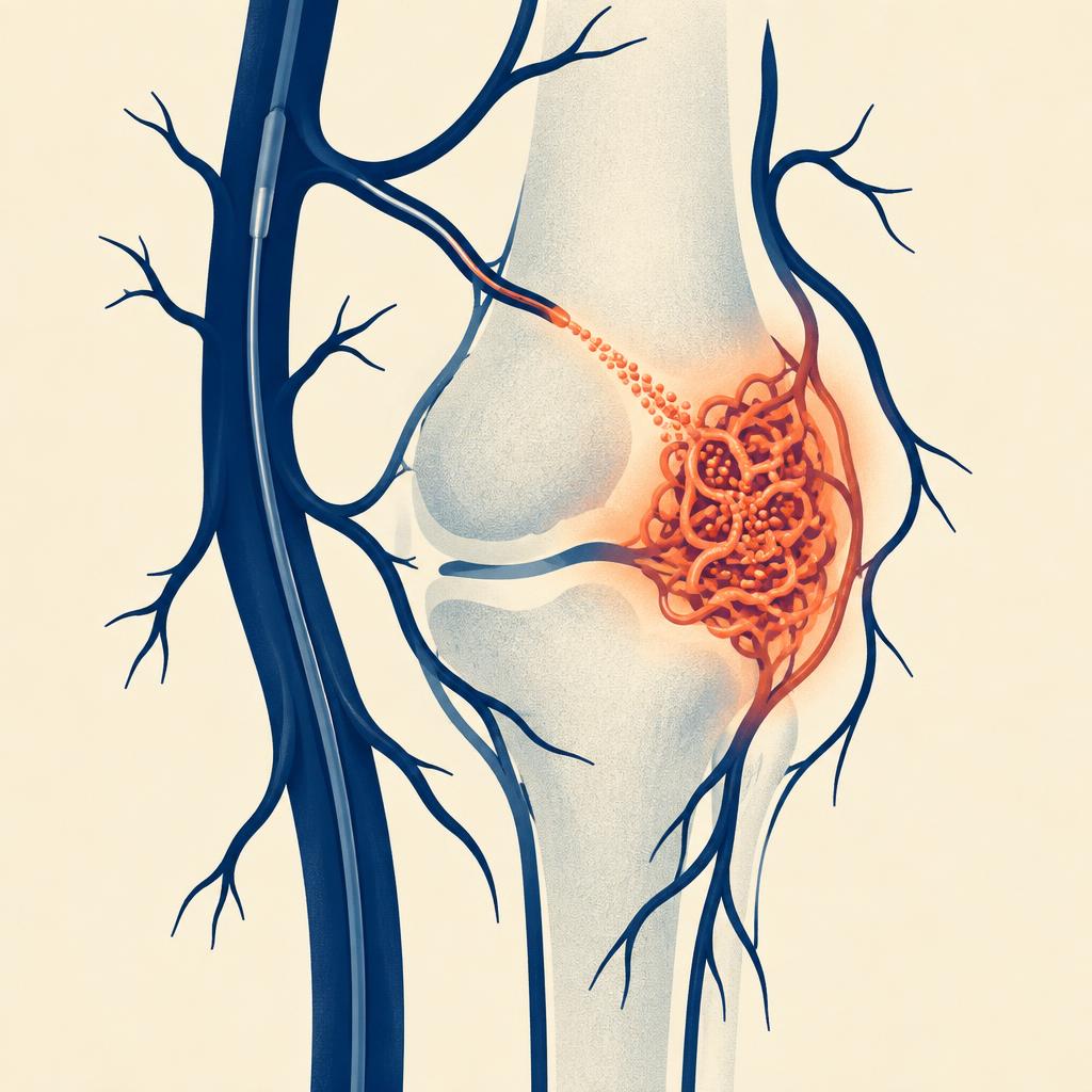
Embolization
A microcatheter delivers microscopic particles into the abnormal branches, reducing blood flow to inflamed synovium.
- 6
Closure & recovery
The catheter is removed and pressure or a closure device seals the access site. You rest for a few hours, then go home.

Recovery timeline
Day 0
Go home the same day with a companion. Keep the access site dry; limit strenuous activity.
Days 1–3
Mild soreness or bruising at the access site. Most people return to light daily activities.
Week 1–2
Some patients notice temporary skin discoloration over the knee; it typically resolves on its own.
Weeks 2–6
Pain relief often begins as inflammation settles. Resume physical therapy and exercise.
Months 1–6
Continued improvement for many patients. Follow-up visit to reassess WOMAC and pain scores.
Before your procedure
- Blood work to check kidney function and clotting.
- Tell your team about blood thinners, diabetes medications, and any contrast dye allergy.
- No food for several hours beforehand, per your care team's instructions.
- Arrange a ride home — you cannot drive after sedation.
Wondering if GAE could help your knee?
Take the 2-minute WOMAC-based candidacy assessment.